
Although at present there is no single imaging method that can be considered ideal based on our review of the literature the standing full-length AP computed radiograph of both lower extremities with the pelvis level along with use of a magnification marker should be the primary imaging modality for the initial evaluation of LLD in the majority of the patients. Diagnosis is made with plain radiographs of the pelvis.

Pelvic X Rays Orthointern
Epos Trade

Posterior Anterior Projection Of Pelvic Radiographs In Children Meets The Correct Positioning Criteria Iranian Journal Of Radiology Full Text
The abdominal radiograph AXR is performed almost exclusively in the supine position and in the AP anteroposterior projection ie.

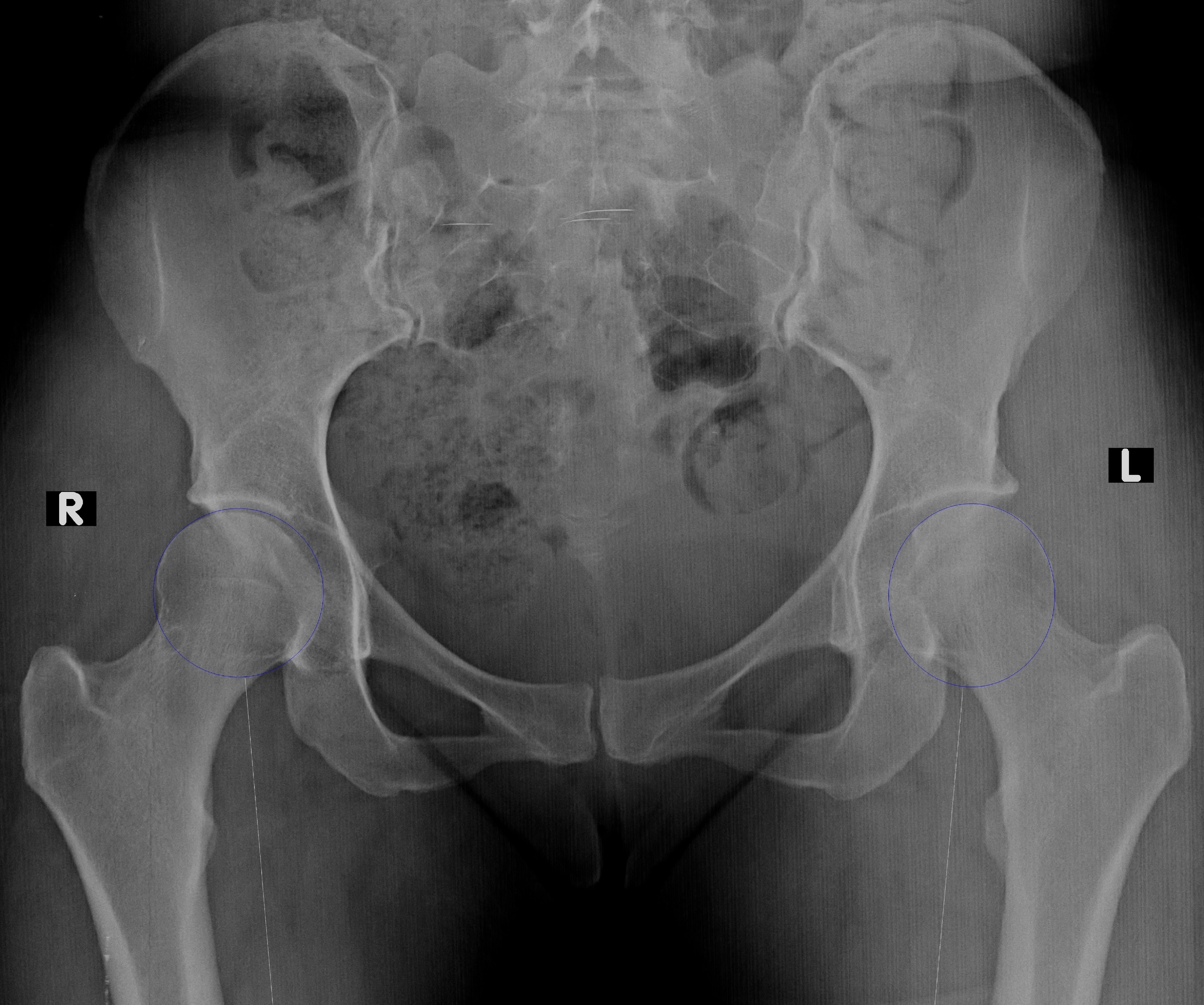
Ap pelvis radiograph. Osteomyelitis is an infection involving bone. Also demonstrates head neck trochanters and proximal one third or one fourth of shaft of femur. Pelvis Fractures in the pediatric population are uncommon injuries that are usually associated with high-energy trauma and are often associated with CNS and abdominal visceral injury.
This article discusses radiographic positioning to show the hip and pelvis for the Radiologic Technologist X-Ray Tech. Osteomyelitis may be classified based on the mechanism of infection hematogenous versus nonhematogenous and the duration of illness acute versus chronic Issues related to the classification epidemiology microbiology clinical manifestations and diagnosis of osteomyelitis in adults are presented here. Radiographic Technique Commonly used radiographic projections of the pelvis and proximal femur include the anteroposterior AP view of the pelvis anterior and posterior oblique Judet views of the pelvis AP view of the hip and frog-leg lateral Dan Miller viewofthehip1-3 TheAPradiographofthepelvisFig1or.
A cross-table lateral radiograph should also be obtained by flexing the unaffected hip and knee and. The AP projection of the abdomen is sometimes called as KUB xray because in this projection the Kidneys Ureters and Bladder are included in the radiograph. It is also use as a preliminary evaluation radiograph or Scot films for some of special procedure.
The chest radiograph also known as the chest x-ray or CXR is anecdotally thought to be the most frequently-performed radiological investigation globally although no published data is known to corroborate thisUK government statistical data from the NHS in England and Wales shows that the chest radiograph remains consistently the most frequently requested imaging test by GPs 2019 dataset 5. Indications This view is of considerable importance in the management of severely injured patients prese. Purpose and Structures Shown Clear image of entire pelvis.
The AP pelvis view is part of a pelvic series examining the iliac crest sacrum proximal femur pubis ischium and the great pelvic ring. The view of the unaffected hip can be used for preoperative planning. When an AP Projection of abdomen taken in supine it is also called as Flat Plate Abdomen.
The first investigation of choice in a patient with hip pain following trauma is a hip x-ray. The anteroposterior AP pelvis view allows the affected and contralateral hips to be compared. Radiograph ESD per radiograph mGy DAP per radiograph Gy cm 2 Abdomen AP.
The anteroposterior AP abdomen projection in contrast includes both halves of the diaphragm. Historically the abdominal radiograph was performed in both supine and erect postures but this practice has been discontinued due to concerns over often excessive radiation dose. Fractures are not always identified on initial x-ray imaging.
This will be an AP radiograph of the pelvis and a lateral radiograph of the painful hip. This is done to ensure that the majority of bowel can be reviewed. If the patient is large more than one film loaded in the Bucky in a landscape direction may be used for each projection.
The x-ray beam passes through the patient from front to back. If there is a clinical concern for a hip fracture but the x-ray is normal further imaging is required. A KUB is a plain frontal supine radiograph of the abdomen.
Radiography Of Pubic Rami Fractures Wikiradiography


Ap Pelvic Radiograph Demonstrating Right Sided Birmingh Open I

Figure Ap Pelvis X Ray Demonstrating A Statpearls Ncbi Bookshelf

Xray Anatomy Of The Hip Medical Radiography Medical Ultrasound Medical Anatomy

Pelvis Ap View Radiology Reference Article Radiopaedia Org
Ap Pelvis Radiograph A And Lateral View B Of A 41 Year Old Woman Download Scientific Diagram

Bubble Sign Ap Pelvis Radiograph Of A Patient With Acute Onset Of Left Download Scientific Diagram
Jbjs Image Quiz Treatment Of Adult Developmental Dysplasia Of The Hip


