
Coli under the Microscope Types Techniques Gram Stain and Hanging Drop Method Introduction E. The results showed that E.

E Coli Bacteria Abc News Australian Broadcasting Corporation

Escherichia Coli E Coli Bacteria Cells Under Microscope Stock Illustration Illustration Of Macrophotography Close 150224499
1
Coli under the microscope.

E coli under microscope. EC E-coli SA S. For example Ecoli O157H7 is one strain causing intestinal infections. It often looks like mucus or a cloudy film over the whole surface of the plate.
Coli B strain with DE3 a λ prophage carrying the T7 RNA polymerase gene and lacI q. The power of a light microscope is limited. Download Full PDF Package.
Not only are SF4 helicases well characterized but E. Coli K12 each has a set of strain-specific genes which make up 26 and 12 of the gene catalogs respectively. Coli DnaB is a homohexameric superfamily 4 SF4 helicase that encircles and translocates along the lagging strand in the 5-3 direction 2021.
A scanning electron microscope SEM is a type of electron microscope that produces images of a sample by scanning the surface with a focused beam of electronsThe electrons interact with atoms in the sample producing various signals that contain information about the surface topography and composition of the sample. Coli cause urinary tract infections. You can detect these rings under a microscope.
In fact MedMD notes 75-95 of UTI infections are caused by E. Coli is a bacterium commonly found in various ecosystems like land and water. 112 Abstract Escherichia coli Bacillus subtilis and Staphylococcus epidermidis were analyzed for this lab activity to determine their Gram Stain.
Coli is a well-tested model organism making this an ideal system for investigating replisome mechanics in vivo. Escherichia coli originally known as Bacterium coli commune was identified in 1885 by the German pediatrician Theodor Escherich 14 29. Download Full PDF Package.
In this type of microscope multiphoton fluorescence excitation results in the capture of high-resolution three-dimensional images of specimen tagged with highly specific. Wet mounts Page 7. Coli is widely distributed in the intestine of.
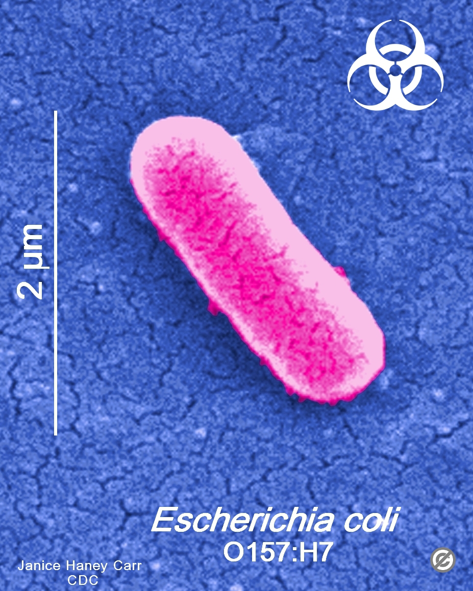
Comma-shaped red cells b. Coli their average size is 15 µm in diameter and 2-6 µm in length. Its even possible to make out structures within the cell such as the nucleus mitochondria and chloroplasts.
Just like Ecoli there are pathogenic strains of S. Coli Salmonella and Shigella have all been found in RTE products. Transformed plasmids containing T7 promoter driven expression are repressed until IPTG induction of T7 RNA polymerase from a.
Coli is commonly studied as they are considered as a standard for the study of different bacteria. Light microscopes use a system of lenses to magnify an image. The most well-known bacteria.
This is substantially more variation than can be tolerated by the species concept as applied to higher organisms and is difficult to reconcile with any definition of species yet devised for microorganisms. Soluble Products of Escherichia coli Induce Mitochondrial Dysfunction-Related Sperm Membrane Lipid Peroxidation Which Is Prevented by Lactobacilli. Your urine will then be examined under a microscope for the presence of bacteria.
B F ompT gal dcm lon hsdS B r B m B λDE3 lacI lacUV5-T7p07 ind1 sam7 nin5 malB K-12 λ S an E. You are examining a drop of aquarium water under the microscope and observe green cells without nuclei. In this type of fluorescence microscope highresolution imaging of thick specimens without physical sectioning can be analyzed using fluorescent-labeled dye.
The electron beam is scanned in a raster scan pattern and the position of. 37 Full PDFs related to this paper. Smaller cells are easily visible under a light microscope.
Escherichia coli E. Other strains of E. A C.
Most of the bacteria range from 02-2 µm in diameter. It teaches the nature of the research process from asking questions and developing a testable hypothesis to writing. Coli Commonly referred to as E.
Coli Escherichia coli is a bacterium that is typically found in a number of environments including various foods soil and animal intestines. Photographs were taken after 24 h incubation at 37 C under 100 magnification using a light microscope indicated over 90 viability with monocytes at 2 4 6 and 24 h of co-culture 24 h photos shown. The CGC has been in operation since 1978 first at the University of Missouri Columbia and since 1992 at the University of Minnesota Twin Cities.
Observation of Bacterial Type I Pili Extension under Fluid Flow. By Doris Stanley July 31 1998. This cellular compartment is found only in those bacteria that have both an outer membrane and plasma membrane eg.
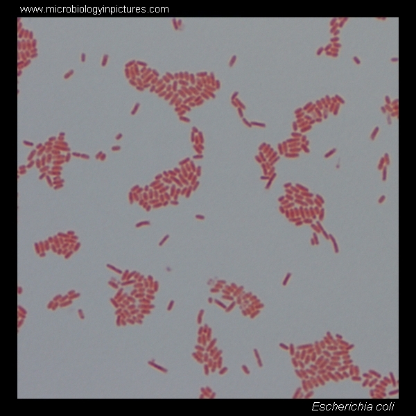
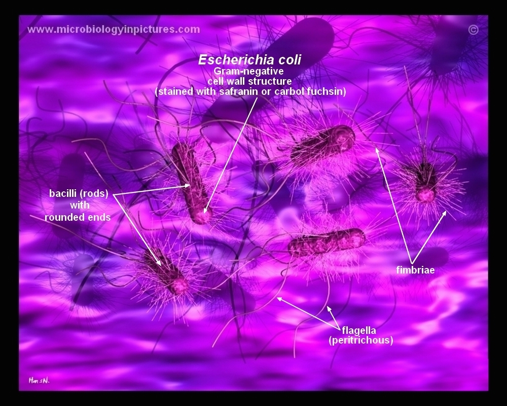
Cooking regimes designed to kill deadly Escherichia coli 0157H7 must be based on the pathogen being in its most heat-resistant state according to a microbiologist with USDAs Agricultural Research ServiceBacteria previously subjected to lower heating temperatures may be tougher to kill. Coli stained pink and classified as Gram-negative. The length can range from 1-10 µm for filamentous or rod-shaped bacteria.
The portion of lipopolysaccharide that is responsible for. Coli can spread to the urinary tract in a variety of ways. In this figure The size comparison between our.
The Caenorhabditis Genetics Center CGC is supported by the National Institutes of Health - Office of Research Infrastructure Programs P40 OD010440. After the multi-layered Gram Stain procedure each bacteria were classified as Gram-positive or Gram-negative depending on their cell walls staining color. Most of the strains of E.
Coli O157H7 and E. Coli Bacteria in Food. The traditional method is based on using cultures examined under a microscope which is time-consuming.
In liquid culture media like Trypticase soy broth or Nutrient broth the growth of the bacterium occurs as a turbidity in the broth medium with a heavy deposits that disperses in the medium on shaking which is further analyzed for the morphology under the microscope gram reaction biochemical tests and Escherichia coli specific tests. Gram negative bacteriaIn the space are enzymes and other proteins that help digest and move nutrients into the cell. The situation therefore is that E.
A method tested in 2005 in a study published in Meat Science is called multiplex PCR. Aureus that are responsible for skin infections abscesses and respiratory infections. Coli is part of the largest group of bacteria called proteobacteria and includes multiple strains.
Coli colony is slightly raised and has an entire fixed margin and a steady growth pattern creating concentric growth rings in the colony. About the CGC. A short summary of this paper.
Proper Heat Treatment Kills Deadly E. Composed of peptidoglycan polysaccharides protein the cell wall maintains the overall shape of a bacterial cell. Coli are harmless but some strains are known to cause diarrhea and even UTIs.
I outline an exercise that can be done easily as part of a microbiology laboratory course. Full PDF Package Download Full PDF Package. The challenge of teaching in the sciences is not only conveying knowledge in the discipline but also developing essential critical thinking data analysis and scientific writing skills.
This bacteria is ideal for the gram staining technique since it is a gram-positive bacteria this means it has a thick peptidoglycan layer that will trap crystal violet and so will appear bluishpurple under the microscope. Coli colony is off-white or beige in color with a shiny texture.
Staphylococcus Aureus And Ecoli Under Microscope Microscopy Of Gram Positive Cocci And Gram Negative Bacilli Morphology And Microscopic Appearance Of Staphylococcus Aureus And E Coli S Aureus Gram Stain And Colony Morphology On Agar Clinical


E Coli Bacteria Preventing E Coli Infections

Escherichia Coli Bacteria E Coli Stock Footage Video 100 Royalty Free 3949175 Shutterstock
Fsa Released Revised E Coli O157 Control Of Cross Contamination Guidance Food Law Latest
Escherichia Coli Light Microscopy

E Coli Gram Stain And Cell Morphology E Coli Micrograph Appearance Under The Microscope E Coli Cell Morphology E Coli Microscopic Picture
Escherichia Coli Smear Prepared Microscope Slide 75x25mm Eisco Labs
How E Coli Bacteria Look Like


